9 0 5 7 8
论文已发表
注册即可获取德孚的最新动态
IF 收录期刊
- 2.6 Breast Cancer (Dove Med Press)
- 3.9 Clin Epidemiol
- 3.3 Cancer Manag Res
- 3.9 Infect Drug Resist
- 3.6 Clin Interv Aging
- 4.8 Drug Des Dev Ther
- 2.8 Int J Chronic Obstr
- 8.0 Int J Nanomed
- 2.3 Int J Women's Health
- 3.2 Neuropsych Dis Treat
- 4.0 OncoTargets Ther
- 2.2 Patient Prefer Adher
- 2.8 Ther Clin Risk Manag
- 2.7 J Pain Res
- 3.3 Diabet Metab Synd Ob
- 4.3 Psychol Res Behav Ma
- 3.4 Nat Sci Sleep
- 1.9 Pharmgenomics Pers Med
- 3.5 Risk Manag Healthc Policy
- 4.5 J Inflamm Res
- 2.3 Int J Gen Med
- 4.1 J Hepatocell Carcinoma
- 3.2 J Asthma Allergy
- 2.3 Clin Cosmet Investig Dermatol
- 3.3 J Multidiscip Healthc

[18F] 氟乙基蟾毒灵 (Bufalin) 在有肝细胞癌的小鼠中的放射合成和药代动力学
Authors Yang Z, Liu J, Huang Q, Zhang Z, Zhang J, Pan Y, Yang Y, Cheng D
Received 10 April 2016
Accepted for publication 20 October 2016
Published 11 January 2017 Volume 2017:10 Pages 329—338
DOI https://doi.org/10.2147/OTT.S110281
Checked for plagiarism Yes
Review by Single-blind
Peer reviewers approved by Dr Chiung-Kuei Huang
Peer reviewer comments 4
Editor who approved publication: Dr Faris Farassati
Purpose: Bufalin, the main component of a Chinese traditional medicine chansu,
shows convincing anticancer effects in a lot of tumor cell lines. However, its
in vivo behavior is still unclear. This research aimed to evaluate how bufalin
was dynamically absorbed after intravenous injection in animal models. We
developed a radiosynthesis method of [18F]fluoroethyl bufalin to noninvasively evaluate the tissue
biodistribution and pharmacokinetics in hepatocellular carcinoma-bearing mice.
Methods: [18F]fluoroethyl bufalin
was synthesized with conjugation of 18F-CH2CH2OTs and bufalin. The radiochemical purity was proved by the
radio-high-performance liquid chromatography (HPLC). The pharmacokinetic
studies of [18F]fluoroethyl bufalin
were then performed in Institute of Cancer Research (ICR) mice. Furthermore,
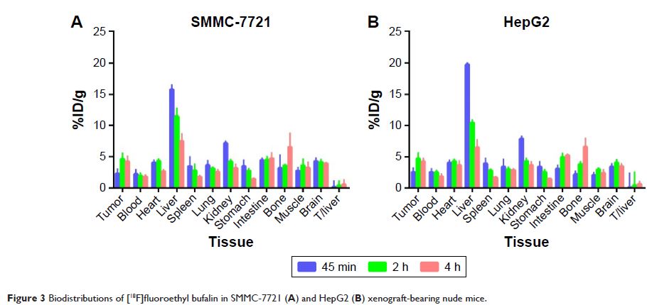
the biodistribution and metabolism of [18F]fluoroethyl bufalin in HepG2 and SMMC-7721 tumor-bearing nude mice
were studied in vivo by micro-positron emission tomography (micro-PET).
Results: The radiochemical purity (RCP) of [18F]fluoroethyl bufalin confirmed by radio-HPLC was 99%±0.18%, and [18F]fluoroethyl bufalin showed good in vitro and in vivo stabilities.
Blood dynamics of [18F]fluoroethyl bufalin conformed to the two compartments in the ICR mice
model. The pharmacokinetic parameters of [18F]fluoroethyl bufalin were calculated by DAS 2.0 software. The area
under concentration–time curve (AUC0–t) and the values of clearance (CL) were 540.137 µg/L·min and
0.001 L/min/kg, respectively. The half-life of distribution (t1/2α ) and
half-life of elimination (t1/2β ) were
0.693 and 510.223 min, respectively. Micro-PET imaging showed that [18F]fluoroethyl bufalin was quickly distributed via the blood circulation;
the major tissue biodistribution of [18F]fluoroethyl bufalin in HepG2 and SMMC-7721 tumor-bearing mice was
liver and bladder.
Conclusion: [18F]fluoroethyl bufalin
was accumulated rapidly in the liver at an early time point (5 min) post
injection (pi) and then declined slowly, mainly through both the hepatic
pathway and the renal pathway. Our study showed the biodistribution of [18F]fluoroethyl bufalin in micro-PET images and provided visible
information for demonstrating the bioactivities of bufalin.
Keywords: [18F]fluoroethyl bufalin,
PET, hepatocellular carcinoma, pharmacokinetic, tissue biodistribution