9 0 5 7 8
论文已发表
注册即可获取德孚的最新动态
IF 收录期刊
- 2.6 Breast Cancer (Dove Med Press)
- 3.9 Clin Epidemiol
- 3.3 Cancer Manag Res
- 3.9 Infect Drug Resist
- 3.6 Clin Interv Aging
- 4.8 Drug Des Dev Ther
- 2.8 Int J Chronic Obstr
- 8.0 Int J Nanomed
- 2.3 Int J Women's Health
- 3.2 Neuropsych Dis Treat
- 4.0 OncoTargets Ther
- 2.2 Patient Prefer Adher
- 2.8 Ther Clin Risk Manag
- 2.7 J Pain Res
- 3.3 Diabet Metab Synd Ob
- 4.3 Psychol Res Behav Ma
- 3.4 Nat Sci Sleep
- 1.9 Pharmgenomics Pers Med
- 3.5 Risk Manag Healthc Policy
- 4.5 J Inflamm Res
- 2.3 Int J Gen Med
- 4.1 J Hepatocell Carcinoma
- 3.2 J Asthma Allergy
- 2.3 Clin Cosmet Investig Dermatol
- 3.3 J Multidiscip Healthc

妊娠期暴露于二氧化钛纳米颗粒会因小鼠中的血管化、增殖和细胞凋亡失调而导致损害胎盘
Authors Zhang L, Xie X, Zhou Y, Yu D, Deng Y, Ouyang J, Yang B, Luo D, Zhang D, Kuang H
Received 23 September 2017
Accepted for publication 11 December 2017
Published 5 February 2018 Volume 2018:13 Pages 777—789
DOI https://doi.org/10.2147/IJN.S152400
Checked for plagiarism Yes
Review by Single-blind
Peer reviewers approved by Dr Alexander Kharlamov
Peer reviewer comments 2
Editor who approved publication: Dr Lei Yang
Background: Titanium dioxide nanoparticles (TiO2 NPs) have
recently found applications in a wide variety of consumer goods. TiO2 NPs exposure significantly increases fetal
deformities and mortality. However, the potential toxicity of TiO2 NPs on the growth and development of placenta has
been rarely studied during mice pregnancy.
Purpose: The objective of this study was to investigate
the effects of maternal exposure of TiO2 NPs on the
placentation.
Methods: Mice were administered TiO2 NPs
by gavage at 0, 1 and 10 mg/kg/day from gestational day (GD) 1 to GD 13. Uteri
and placentas from these mice were collected and counted the numbers of
implanted and resorbed embryo and measured the placental weight on GD 13.
Placental morphometry was observed by hematoxylin and eosin staining. The levels
of Hand1 , Esx1 , Eomes , Hand2 , Ascl2 and Fra1 mRNA were assessed by
qRT-PCR. Uterine NK (uNK) cells were detected by using DBA lectin. Laminin
immunohistochemical staining was to identify fetal vessels. Western blotting
and transmission electron micrograph (TEM) were used to assess the apoptosis of
placenta.
Results: No treatment-related difference was observed in
the numbers of implanted and resorbed embryos and weight of placenta between
the groups. However, 1 mg/kg/day TiO2 NPs
treatment significantly reduced the ratio of placenta/body weight on GD 13. The
proportion of spongiotrophoblast in the 10 mg/kg/day dose group became higher
than that in the control group, yet that of labyrinth was significantly lower
in 10 mg/kg/day mice. The expression levels of Hand1 , Esx1 , Eomes , Hand2 , Ascl2 and Fra1 mRNA markedly
decreased in TiO2 NP treated placentas.
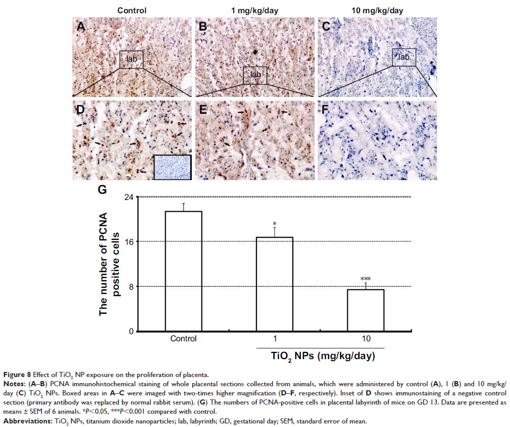
Furthermore, TiO2 NPs treatment impaired the
formation of intricate networks of fetal vessels and reduced the number of uNK
cells, and inhibited proliferation and induced apoptosis of placenta by nuclear
pyknosis, the activation of caspase-3 and upregulation of Bax protein and
downregulation of Bcl-2 protein on GD 13.
Conclusion: Gestational exposure to TiO2 NPs significantly impairs the growth and
development of placenta in mice, with a mechanism that seems to be involved in
the dysregulation of vascularization, proliferation and apoptosis. Therefore,
our results suggested the need for great caution while handling of the
nanomaterials by workers and specially pregnant consumers.
Keywords: nanoparticles,
titanium dioxide, placenta, proliferation, apoptosis, vascularization