9 0 6 7 6
论文已发表
注册即可获取德孚的最新动态
IF 收录期刊
- 2.6 Breast Cancer (Dove Med Press)
- 3.9 Clin Epidemiol
- 3.3 Cancer Manag Res
- 3.9 Infect Drug Resist
- 3.6 Clin Interv Aging
- 4.8 Drug Des Dev Ther
- 2.8 Int J Chronic Obstr
- 8.0 Int J Nanomed
- 2.3 Int J Women's Health
- 3.2 Neuropsych Dis Treat
- 4.0 OncoTargets Ther
- 2.2 Patient Prefer Adher
- 2.8 Ther Clin Risk Manag
- 2.7 J Pain Res
- 3.3 Diabet Metab Synd Ob
- 4.3 Psychol Res Behav Ma
- 3.4 Nat Sci Sleep
- 1.9 Pharmgenomics Pers Med
- 3.5 Risk Manag Healthc Policy
- 4.5 J Inflamm Res
- 2.3 Int J Gen Med
- 4.1 J Hepatocell Carcinoma
- 3.2 J Asthma Allergy
- 2.3 Clin Cosmet Investig Dermatol
- 3.3 J Multidiscip Healthc

CT 切片厚度对胸癌放疗中体积和剂量评估的影响
Authors Luo H, He Y, Jin F, Yang D, Liu X, Ran X, Wang Y
Received 15 May 2018
Accepted for publication 26 July 2018
Published 20 September 2018 Volume 2018:10 Pages 3679—3686
DOI https://doi.org/10.2147/CMAR.S174240
Checked for plagiarism Yes
Review by Single-blind
Peer reviewers approved by Dr Cristina Weinberg
Peer reviewer comments 3
Editor who approved publication: Dr Antonella D'Anneo
Introduction: Accurate delineation of targets and organs at risk (OAR) is
required to ensure treatment efficacy and minimize risk of normal tissue
toxicity with radiotherapy. Therefore, we evaluated the impacts of computed
tomography (CT) slice thickness and reconstruction methods on the volume and
dose evaluations of targets and OAR.
Patients and
methods: Eleven CT datasets from patients
with thoracic cancer were included. 3D images with a slice thickness of 2 mm
(2–CT) were created automatically. Images of other slice thickness (4–CT, 6–CT,
8–CT, 10–CT) were reconstructed manually by the selected 2D images using two
methods; internal tumor information and external CT Reference markers.
Structures and plans on 2–CT images, as a reference data, were copied to the
reconstructed images.
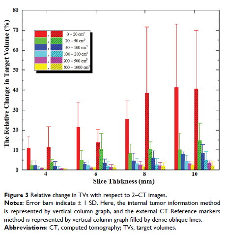
Results: The maximum error of volume was 84.6% for the smallest target in
10–CT, and the maximum error (≥20 cm3) was 10.1%, 14.8% for the two reconstruction methods, internal tumor
information and external CT Reference, respectively. Changes in conformity
index for a target of <20 cm3 were 5.4% and 17.5% in 8–CT. Changes on V30 and V40 of the heart were considerable. In the internal tumor information
method, volumes of hearts decreased by 3.2% in 6–CT, while V30 and V40 increased by 18.4% and 46.6%.
Conclusion: The image reconstruction method by internal tumor information was
less affected by slice thickness than the image reconstruction method by
external CT Reference markers. This study suggested that before positioning
scanning, the largest section through the target should be determined and the
optimal slice thickness should be estimated.
Keywords: computed tomography, slice thickness, thoracic cancer, dose, image
reconstruction
