9 0 5 7 8
论文已发表
注册即可获取德孚的最新动态
IF 收录期刊
- 2.6 Breast Cancer (Dove Med Press)
- 3.9 Clin Epidemiol
- 3.3 Cancer Manag Res
- 3.9 Infect Drug Resist
- 3.6 Clin Interv Aging
- 4.8 Drug Des Dev Ther
- 2.8 Int J Chronic Obstr
- 8.0 Int J Nanomed
- 2.3 Int J Women's Health
- 3.2 Neuropsych Dis Treat
- 4.0 OncoTargets Ther
- 2.2 Patient Prefer Adher
- 2.8 Ther Clin Risk Manag
- 2.7 J Pain Res
- 3.3 Diabet Metab Synd Ob
- 4.3 Psychol Res Behav Ma
- 3.4 Nat Sci Sleep
- 1.9 Pharmgenomics Pers Med
- 3.5 Risk Manag Healthc Policy
- 4.5 J Inflamm Res
- 2.3 Int J Gen Med
- 4.1 J Hepatocell Carcinoma
- 3.2 J Asthma Allergy
- 2.3 Clin Cosmet Investig Dermatol
- 3.3 J Multidiscip Healthc

羟基磷灰石纳米粒子的体外和体内抗黑素瘤作用:物质因素的影响
Authors Wu H, Li Z, Tang J, Yang X, Zhou Y, Guo B, Wang L, Zhu X, Tu C, Zhang X
Received 1 October 2018
Accepted for publication 16 January 2019
Published 15 February 2019 Volume 2019:14 Pages 1177—1191
DOI https://doi.org/10.2147/IJN.S184792
Checked for plagiarism Yes
Review by Single-blind
Peer reviewers approved by Dr Thiruganesh Ramasamy
Peer reviewer comments 4
Editor who approved publication: Dr Lei Yang
Background: Treatment
for melanoma is a challenging clinical problem, and some new strategies are
worth exploring.
Purpose: The
objective of this study was to investigate the in vitro and in vivo
anti-melanoma effects of hydroxyapatite nanoparticles (HANPs) and discuss the
involved material factors.
Materials and methods: Five
types of HANPs, ie, HA-A, HA-B, HA-C, HA-D, and HA-E, were prepared by wet
chemical method combining with polymer template and appropriate
post-treatments. The in vitro effects of the as-prepared five HANPs on
inhibiting the viability of A375 melanoma cells and inducing the apoptosis of
the cells were evaluated by Cell Counting Kit-8 analysis, cell nucleus
morphology observation, flow cytometer, and PCR analysis. The in vivo
anti-melanoma effects of HANPs were studied in the tumor model of nude mice.
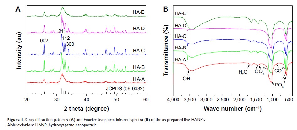
Results: The five
HANPs had different physicochemical properties, including morphology, size,
specific surface area (SSA), crystallinity, and so on. By the in vitro cell
study, it was found that the material factors played important roles in the
anti-melanoma effect of HANPs. Among the as-prepared five HANPs, HA-A with
granular shape, smaller size, higher SSA, and lower crystallinity exhibited
best effect on inhibiting the viability of A375 cells. At the concentration of
200 µg/mL, HA-A resulted in the lowest cell viability (34.90%) at day 3. All
the HANPs could induce the apoptosis of A375 cells, and the relatively higher
apoptosis rates of the cells were found in HA-A (20.10%) and HA-B (19.41%) at
day 3. However, all the HANPs showed no inhibitory effect on the viability of
the normal human epidermal fibroblasts. The preliminary in vivo evaluation
showed that both HA-A and HA-C could delay the formation and growth speed of
melanoma tissue significantly. Likely, HA-A exhibited better effect on
inhibiting the growth of melanoma tissue than HA-C. The inhibition rate of HA-A
for tumor tissue growth reached 49.1% at day 23.
Conclusion: The
current study confirmed the anti-melanoma effect of HANPs and provided a new
idea for the clinical treatment of melanoma.
Keywords: hydroxyapatite,
nanoparticles, melanoma cells, fibroblasts, viability, apoptosis, tumor,
suppression
