9 0 5 7 8
论文已发表
注册即可获取德孚的最新动态
IF 收录期刊
- 2.6 Breast Cancer (Dove Med Press)
- 3.9 Clin Epidemiol
- 3.3 Cancer Manag Res
- 3.9 Infect Drug Resist
- 3.6 Clin Interv Aging
- 4.8 Drug Des Dev Ther
- 2.8 Int J Chronic Obstr
- 8.0 Int J Nanomed
- 2.3 Int J Women's Health
- 3.2 Neuropsych Dis Treat
- 4.0 OncoTargets Ther
- 2.2 Patient Prefer Adher
- 2.8 Ther Clin Risk Manag
- 2.7 J Pain Res
- 3.3 Diabet Metab Synd Ob
- 4.3 Psychol Res Behav Ma
- 3.4 Nat Sci Sleep
- 1.9 Pharmgenomics Pers Med
- 3.5 Risk Manag Healthc Policy
- 4.5 J Inflamm Res
- 2.3 Int J Gen Med
- 4.1 J Hepatocell Carcinoma
- 3.2 J Asthma Allergy
- 2.3 Clin Cosmet Investig Dermatol
- 3.3 J Multidiscip Healthc

非呼吸暂停相关的睡眠破碎对脑血管病患者认知功能的影响
Authors Wang J, Chen X, Liao J, Zhou L, Liao S, Shan Y, Lu Z, Tao J
Received 8 November 2018
Accepted for publication 14 February 2019
Published 18 April 2019 Volume 2019:15 Pages 1009—1014
DOI https://doi.org/10.2147/NDT.S193869
Checked for plagiarism Yes
Review by Single-blind
Peer reviewers approved by Dr Colin Mak
Peer reviewer comments 2
Editor who approved publication: Dr Jun Chen
Background: Cognitive
impairment in patients with cerebral small vessel disease (CSVD) is common, but
the pathogenic mechanism is not well understood. The situation of
non-breathing-related sleep fragmentation in CSVD patients and its influence on
cognitive impairment is not clear. The aim of this study was to investigate the
influence of non-breathing-related sleep fragmentation on cognitive function in
patients with CSVD.
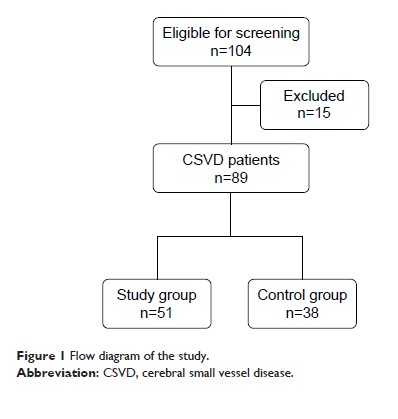
Methods: A group
of 89 CSVD patients without breathing-related sleep disorders in the Department
of Neurology, Third Affiliated Hospital of Sun Yat-sen University was enrolled.
The patients underwent magnetic resonance scan, polysomnography, cognitive
function evaluation using Montreal Cognitive Assessment scale (MoCA), and
Mini-Mental State Examination. The patients were assigned to study group
(arousal index [ArI] ≥26.8/hour) or control group (ArI <26.8/hour) based on
the average level of ArI (mean =26.8, SD =7.5) at night, and the cognitive
function of the patients in the two groups was analyzed.
Results: The total
MoCA score, the subscale scores of visuospatial ability and delayed recall in
the study group were significantly lower than that in the control group (P <0.05). The
cognitive impairment measured by MoCA was positively related to ArI level and
%N-3 sleep according to the results of logistic regression (P <0.05).
Conclusion: Non-breathing-related
sleep fragmentation is associated with cognitive impairment in CSVD patients,
especially executive function and delayed recall ability.
Keywords: cerebrovascular
disorders, sleep, fragmentation, cognition, polysomnography
