9 0 6 7 6
论文已发表
注册即可获取德孚的最新动态
IF 收录期刊
- 2.6 Breast Cancer (Dove Med Press)
- 3.9 Clin Epidemiol
- 3.3 Cancer Manag Res
- 3.9 Infect Drug Resist
- 3.6 Clin Interv Aging
- 4.8 Drug Des Dev Ther
- 2.8 Int J Chronic Obstr
- 8.0 Int J Nanomed
- 2.3 Int J Women's Health
- 3.2 Neuropsych Dis Treat
- 4.0 OncoTargets Ther
- 2.2 Patient Prefer Adher
- 2.8 Ther Clin Risk Manag
- 2.7 J Pain Res
- 3.3 Diabet Metab Synd Ob
- 4.3 Psychol Res Behav Ma
- 3.4 Nat Sci Sleep
- 1.9 Pharmgenomics Pers Med
- 3.5 Risk Manag Healthc Policy
- 4.5 J Inflamm Res
- 2.3 Int J Gen Med
- 4.1 J Hepatocell Carcinoma
- 3.2 J Asthma Allergy
- 2.3 Clin Cosmet Investig Dermatol
- 3.3 J Multidiscip Healthc

Management of progressive keratoconus with partial topography-guided PRK combined with refractive, customized CXL – a novel technique: the enhanced Athens protocol
Authors Kanellopoulos AJ
Received 24 September 2018
Accepted for publication 19 December 2018
Published 2 April 2019 Volume 2019:13 Pages 581—588
DOI https://doi.org/10.2147/OPTH.S188517
Checked for plagiarism Yes
Review by Single-blind
Peer reviewers approved by Dr Cristina Weinberg
Peer reviewer comments 2
Editor who approved publication: Dr Scott Fraser
Purpose: To report a novel application of partial
topography-guided photorefractive keratectomy combined with topographically
customized, higher fluence, and variable pattern corneal cross-linking applied
on the same day of the treatment of keratoconus.
Methods: A
topography-guided partial photorefractive keratectomy treatment of maximum
30 mm over the thinnest cone area was applied initially followed by a
7 mm, 50 mm phototherapeutic keratectomy treatment to address
epithelial removal. 0.02% Mitomycin C was applied for 20 seconds and then
the exposed stroma was soaked with 0.1% riboflavin solution for 5 minutes.
The cornea was then treated with a customized, variable-pattern and
20 mW/cm2 fluence for a total of 5–10 J, and up to 15 J of energy was
delivered with the KXL-II device employing an active tracker. The center of the
pattern that received the 15 J was topography-matched with the thinnest area of
the cone. Visual acuity, refractive error, cornea clarity, keratometry,
topography, pachymetry with a multitude of modalities and endothelial cell
density were evaluated over 36 months.
Results: Keratoconus
was stabilized in all cases. The severity of keratoconus stage by
Amsler–Krumeich criteria improved from an average of 3.2 (1–4) to 1.8 (0–3).
Uncorrected distance visual acuity changed from preoperative 20/80 to 20/25 at
6 months. A maximum astigmatic reduction of 7.8 D (5.3–15.6), and a significant
cornea surface normalization (an index of height decentration improvement from
0.155 [±0.065] to 0.045 [±0.042]) were achieved by 1 month and remained
relatively stable for 36 months postoperatively. Two cases delayed full
reepithelialization for up to 9 days.
Conclusion: This
paper introduces a novel technique in order to maximize the refractive
normalization effect along with ectasia stabilization in young keratoconus
patients. This may facilitate the use of less tissue ablation, in comparison to
utilizing a homogeneous UV light beam for corneal cross-linking in Athens
Protocol cases. It broadens the number of potential candidate cases that would
have been limited to employ this technique due to tissue thickness limitations.
Keywords: corneal
ectasia, corneal irregularity-normalization, therapeutic excimer ablation
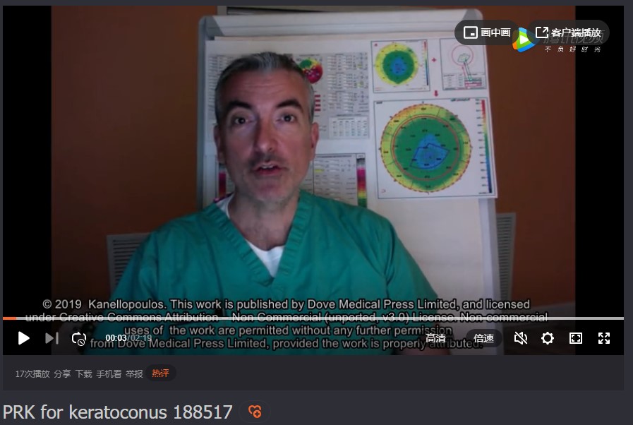
摘要视频链接:PRK for keratoconus