9 0 9 6 8
论文已发表
注册即可获取德孚的最新动态
IF 收录期刊
- 2.6 Breast Cancer (Dove Med Press)
- 3.9 Clin Epidemiol
- 3.3 Cancer Manag Res
- 3.9 Infect Drug Resist
- 3.6 Clin Interv Aging
- 4.8 Drug Des Dev Ther
- 2.8 Int J Chronic Obstr
- 8.0 Int J Nanomed
- 2.3 Int J Women's Health
- 3.2 Neuropsych Dis Treat
- 4.0 OncoTargets Ther
- 2.2 Patient Prefer Adher
- 2.8 Ther Clin Risk Manag
- 2.7 J Pain Res
- 3.3 Diabet Metab Synd Ob
- 4.3 Psychol Res Behav Ma
- 3.4 Nat Sci Sleep
- 1.9 Pharmgenomics Pers Med
- 3.5 Risk Manag Healthc Policy
- 4.5 J Inflamm Res
- 2.3 Int J Gen Med
- 4.1 J Hepatocell Carcinoma
- 3.2 J Asthma Allergy
- 2.3 Clin Cosmet Investig Dermatol
- 3.3 J Multidiscip Healthc

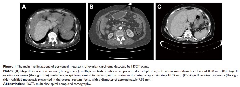
多层螺旋 CT 和磁共振成像对原发性卵巢癌腹膜转移诊断的比较
Authors Guo HL, He L, Zhu YC, Wu K, Yuan F
Received 29 July 2017
Accepted for publication 15 September 2017
Published 28 February 2018 Volume 2018:11 Pages 1087—1094
DOI https://doi.org/10.2147/OTT.S147700
Checked for plagiarism Yes
Review by Single-blind
Peer reviewers approved by Dr Jia Fan
Peer reviewer comments 2
Editor who approved publication: Dr William Cho
Abstract: The advent of disease evaluation by means of multi-slice spiral
computed tomography (MSCT) and magnetic resonance imaging (MRI) represents a
continually emerging role in the evaluation of various diseases; however, its
role is yet to be adequately defined. Thus, the aim of the study was to compare
the diagnostic value of MSCT and MRI in the diagnosis of peritoneal metastasis
in primary ovarian carcinoma. Between January 2013 and December 2015, MSCT or
MRI data were collected from 42 patients who had been previously diagnosed with
peritoneal metastasis of ovarian carcinoma at the First Affiliated Hospital of
Kunming Medical University. The tumor location, size, edge, and shape were all
evaluated independently by three qualified imaging physicians using a
double-blind method to confirm whether the patients were indeed suffering from
peritoneal metastasis, as well as to rank the metastatic lesions recorded on a
five-point scale. It was hypothesized that MRI and MSCT were comparable in the
evaluation of ovarian carcinoma. Therefore, a receiver operating
characteristics (ROC) curve was used to analyze the results and also to
directly compare the respective diagnostic values of MSCT and MRI. In total,
165 metastatic lesions were confirmed by means of surgical operation. MSCT
revealed 131 metastatic lesions, while MRI confirmed 154 metastatic lesions.
The metastatic sites were primarily located on the subphrenic, epiploon, and
gastrocolic ligaments and were further confirmed by either MRI or CT. In regard
to MSCT, the most common site of underdiagnoses was in the vicinity of the
uterus–rectum–fossa. MRI displayed a high detection rate in every site. The
omission diagnostic rate of MSCT and MRI were 20.61% and 6.67%, respectively,
while the accuracy rates were 79.39% and 93.33%, respectively. The obtained
results revealed that the MSCT value of area under the ROC curve was smaller
than that for MRI. Our findings provided evidence asserting that MRI, in
comparison to MSCT, was more accurate in diagnosing peritoneal metastasis in
patients with ovarian carcinoma.
Keywords: multi-slice spiral computed tomography, magnetic resonance
imaging, ovarian carcinoma, peritoneal metastasis, accuracy, omission
diagnostic rate, ROC curves