9 0 8 0 2
论文已发表
注册即可获取德孚的最新动态
IF 收录期刊
- 2.6 Breast Cancer (Dove Med Press)
- 3.9 Clin Epidemiol
- 3.3 Cancer Manag Res
- 3.9 Infect Drug Resist
- 3.6 Clin Interv Aging
- 4.8 Drug Des Dev Ther
- 2.8 Int J Chronic Obstr
- 8.0 Int J Nanomed
- 2.3 Int J Women's Health
- 3.2 Neuropsych Dis Treat
- 4.0 OncoTargets Ther
- 2.2 Patient Prefer Adher
- 2.8 Ther Clin Risk Manag
- 2.7 J Pain Res
- 3.3 Diabet Metab Synd Ob
- 4.3 Psychol Res Behav Ma
- 3.4 Nat Sci Sleep
- 1.9 Pharmgenomics Pers Med
- 3.5 Risk Manag Healthc Policy
- 4.5 J Inflamm Res
- 2.3 Int J Gen Med
- 4.1 J Hepatocell Carcinoma
- 3.2 J Asthma Allergy
- 2.3 Clin Cosmet Investig Dermatol
- 3.3 J Multidiscip Healthc

细胞质 TIMP3 与 miR-222 之间的负相关性表明 NSCLC 预后良好
Authors Lei Y, Liu Z, Yang W
Received 28 April 2018
Accepted for publication 10 July 2018
Published 6 September 2018 Volume 2018:11 Pages 5551—5557
DOI https://doi.org/10.2147/OTT.S172522
Checked for plagiarism Yes
Review by Single-blind
Peer reviewers approved by Dr Cristina Weinberg
Peer reviewer comments 2
Editor who approved publication: Dr Sanjeev Srivastava
Background: The aim of this study was to observe the expression of microRNA-222 (miR-222) and matrix metalloproteinase inhibitor 3 (TIMP3) in non-small cell lung cancer (NSCLC) and discuss their significance.
Methods: A total of 230 patients with NSCLC were enrolled in the observation group during the operation. Ninety-eight normal adjacent tissues were used as the control group. Two groups of miR-222 and TIMP3 were detected by in situ hybridization and immunohistochemistry. The distribution of miR-222 and TIMP3 in A549/H358/PC9 cells was observed by immunofluorescence. Chi-squared and Spearman correlation tests were used to analyze the relationship among miR-222, TIMP3 expression, and clinicopathological parameters of NSCLC. Kaplan–Meier and Cox proportional hazards regression were used to analyze the prognostic impact of miR-222 and TIMP3.
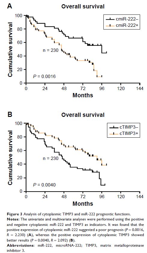
Results: Immunohistochemistry showed that the expression of miR-222 in lung cancer tissue was significantly higher, but TIMP3 was lower than that in normal lung tissue (P = 0.0001 for the former and P = 0.0002 for the latter). Meanwhile, miR-222 and TIMP3 were mainly distributed in the cytoplasm. Among them, cTIMP3 accounted for 70.29% (72/101), cmiR-222 for 59.35% (92/155), 14.85% for nTIMP3 (15/101), and 18.06% for nmiR-222 (28/155). There was a significant difference in distribution (both P < 0.0001). The expression of miR-222 and TIMP3 were negatively correlated in lung cancer tissues (r = -0.43, P = 0.0219). With the progression of clinical stage, the positive intensity of cTIMP3 showed a decreasing trend, while the cmiR-222 showed a reverse trend (the former P = 0.0024 and the latter P < 0.0001). In the Kaplan–Meier prognostic analysis, we found that the high expression of cTIMP3 could predict a better prognosis (P = 0.0040), whereas cmiR-222 was the opposite (P = 0.0016). Multivariate analysis shows that both can be used as independent factors.
Conclusion: TIMP3 expression in lung cancer is relatively low and has a negative correlation with lung cancer staging and prognosis, suggesting that it may play a defensive function in the development of lung cancer, while miR-222 has the opposite effect, and the expression of both proteins is negatively correlated, suggesting that in lung cancer progresses, both proteins may play some role together.
Keywords: lung cancer,tumor markers, A549/h358/pc9, biological mechanism, immuno-fluorescence
