9 0 8 1 0
论文已发表
注册即可获取德孚的最新动态
IF 收录期刊
- 2.6 Breast Cancer (Dove Med Press)
- 3.9 Clin Epidemiol
- 3.3 Cancer Manag Res
- 3.9 Infect Drug Resist
- 3.6 Clin Interv Aging
- 4.8 Drug Des Dev Ther
- 2.8 Int J Chronic Obstr
- 8.0 Int J Nanomed
- 2.3 Int J Women's Health
- 3.2 Neuropsych Dis Treat
- 4.0 OncoTargets Ther
- 2.2 Patient Prefer Adher
- 2.8 Ther Clin Risk Manag
- 2.7 J Pain Res
- 3.3 Diabet Metab Synd Ob
- 4.3 Psychol Res Behav Ma
- 3.4 Nat Sci Sleep
- 1.9 Pharmgenomics Pers Med
- 3.5 Risk Manag Healthc Policy
- 4.5 J Inflamm Res
- 2.3 Int J Gen Med
- 4.1 J Hepatocell Carcinoma
- 3.2 J Asthma Allergy
- 2.3 Clin Cosmet Investig Dermatol
- 3.3 J Multidiscip Healthc

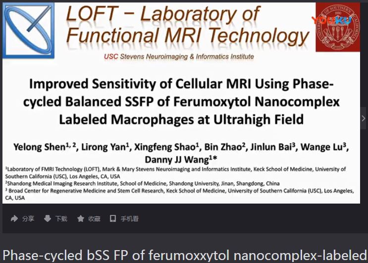
Improved sensitivity of cellular MRI using phase-cycled balanced SSFP of ferumoxytol nanocomplex-labeled macrophages at ultrahigh field
Authors Shen Y, Yan L, Shao X, Zhao B, Bai J, Lu W, Wang DJJ
Received 1 April 2018
Accepted for publication 4 May 2018
Published 3 July 2018 Volume 2018:13 Pages 3839—3852
DOI https://doi.org/10.2147/IJN.S169860
Checked for plagiarism Yes
Review by Single-blind
Peer reviewers approved by Dr Thiruganesh Ramasamy
Peer reviewer comments 3
Editor who approved publication: Prof. Dr. Thomas J Webster
Purpose: The purpose of this study was to investigate the feasibility and
sensitivity of cellular magnetic resonance imaging (MRI) with ferumoxytol
nanocomplex-labeled macrophages at ultrahigh magnetic field of 7 T.
Materials and methods: THP-1-induced macrophages were labeled using
self-assembling heparin + protamine + ferumoxytol nanocomplexes which were
injected into a gelatin phantom visible on both microscope and MRI.
Susceptibility-weighted imaging (SWI) and balanced steady-state free precession
(bSSFP) pulse sequences were applied at 3 and 7 T. The average, maximum
intensity projection, and root mean square combined images were generated for
phase-cycled bSSFP images. The signal-to-noise ratio and contrast-to-noise
ratio (CNR) efficiencies were calculated. Ex vivo experiments were then
performed using a formalin-fixed pig brain injected with ~100 and ~1,000
labeled cells, respectively, at both 3 and 7 T.
Results: A high cell labeling efficiency (>90%) was
achieved with heparin + protamine + ferumoxytol nanocomplexes. Less than 100
cells were detectable in the gelatin phantom at both 3 and 7 T. The 7 T data
showed more than double CNR efficiency compared to the corresponding sequences
at 3 T. The CNR efficiencies of phase-cycled bSSFP images were higher compared to
those of SWI, and the root mean square combined bSSFP showed the highest CNR
efficiency with minimal banding. Following co-registration of microscope and MR
images, more cells (51/63) were detected by bSSFP at 7 T than at 3 T (36/63).
On pig brain, both ~100 and ~1,000 cells were detected at 3 and 7 T. While the
cell size appeared larger due to blooming effects on SWI, bSSFP allowed better
contrast to precisely identify the location of the cells with higher
signal-to-noise ratio efficiency.
Conclusion: The proposed cellular MRI with ferumoxytol
nanocomplex-labeled macrophages at 7 T has a high sensitivity to detect <100
cells. The proposed method has great translational potential and may have broad
clinical applications that involve cell types with a primary phagocytic
phenotype.
Keywords: ultrasmall
superparamagnetic iron oxide nanoparticles, ultrahigh field, balanced
steady-state free precession, cellular magnetic resonance imaging,
self-assembling nanocomplexes, 7 T
摘要视频链接:Phase-cycled bSSFP of
ferumoxxytol nanocomplex-labeled macrophages at 7T
