115195
论文已发表
注册即可获取德孚的最新动态
IF 收录期刊
- 3.6 Breast Cancer (Dove Med Press)
- 4.3 Clin Epidemiol
- 2.6 Cancer Manag Res
- 3.2 Infect Drug Resist
- 4.1 Clin Interv Aging
- 6.1 Drug Des Dev Ther
- 4.1 Int J Chronic Obstr
- 8.7 Int J Nanomed
- 2.5 Int J Women's Health
- 3.2 Neuropsych Dis Treat
- 2.4 OncoTargets Ther
- 2.6 Patient Prefer Adher
- 2.6 Ther Clin Risk Manag
- 3.1 J Pain Res
- 3.5 Diabet Metab Synd Ob
- 4.5 Psychol Res Behav Ma
- 3.4 Nat Sci Sleep
- 2.4 Pharmgenomics Pers Med
- 2.6 Risk Manag Healthc Policy
- 4.6 J Inflamm Res
- 2.3 Int J Gen Med
- 3.9 J Hepatocell Carcinoma
- 3.3 J Asthma Allergy
- 2.5 Clin Cosmet Investig Dermatol
- 3.0 J Multidiscip Healthc

CT 扫描在结直肠癌分类中的作用
Authors Li ZH, You DY, Gao DP, Yang GJ, Dong XX, Zhang DF, Ding YY
Received 23 December 2016
Accepted for publication 23 March 2017
Published 26 April 2017 Volume 2017:10 Pages 2297—2303
DOI https://doi.org/10.2147/OTT.S131008
Checked for plagiarism Yes
Review by Single-blind
Peer reviewers approved by Dr Ru Chen
Peer reviewer comments 4
Editor who approved publication: Dr William Cho
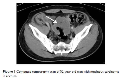
Objectives: Most colorectal cancers are classical adenocarcinomas (AC), and less frequent
subtypes include mucinous adenocarcinomas (MAC) and signet-ring cell carcinomas
(SC). The purpose of this study was to evaluate the computed tomography (CT)
findings that can help to differentiate MAC and SC from AC.
Methods: CT scans of 168 patients with pathologically proven
MAC and 67 patients with pathologically proven SC were analyzed, and 220
patients with classical AC were also included as a control group. CT findings
of the three groups were compared and contrasted in terms of the bowel involvement
patterns, contrast enhancement patterns, and presence or absence of bowel
obstruction, intratumoral calcification, pericolic fat infiltration, and local
tumor extension to adjacent organs. Statistical analyses were made by using the
one-way analysis of variance, least significant difference test, and Pearson’s
chi-square test.
Results: Compared with classical AC, the MAC showed more severe
(6.29±2.69 cm vs 4.57±1.74 cm, P <0.001) and higher percentage
of occurrence of eccentric bowel-wall thickening (37.2% vs 11.5%, P <0.001).
Heterogeneous contrast enhancement was most common in MAC (P <0.01), and MAC showed more
areas with hypoattenuation (P <0.001). The
presence of intratumoral calcification was most frequent in MAC (17.9% vs 2% vs
6.8%) (P <0.001); the SC also were more
severe (5.75±2.28 cm vs 4.57±1.74 cm. P =0.001) than AC, but SC tend to
show more cases of concentric even bowel-wall thickening (67.2%); homogeneous
contrast enhancement was most common in SC (P <0.01), and it
showed a target appearance. The presence of peritoneal seeding was most
frequent in SC (35.8% vs 8% vs 2.7%, P <0.001), while the presence of
regional lymph node metastasis (P =0.190) and direct
invasion of adjacent organs or metastasis (P =0.323) were not
significantly different among them.
Conclusion: Some radiological features by CT can be used to
classify different colon tumor types.
Keywords: cancer, colorectal, mucinous,
prognosis, signet ring, computed tomography