115195
论文已发表
注册即可获取德孚的最新动态
IF 收录期刊
- 3.6 Breast Cancer (Dove Med Press)
- 4.3 Clin Epidemiol
- 2.6 Cancer Manag Res
- 3.2 Infect Drug Resist
- 4.1 Clin Interv Aging
- 6.1 Drug Des Dev Ther
- 4.1 Int J Chronic Obstr
- 8.7 Int J Nanomed
- 2.5 Int J Women's Health
- 3.2 Neuropsych Dis Treat
- 2.4 OncoTargets Ther
- 2.6 Patient Prefer Adher
- 2.6 Ther Clin Risk Manag
- 3.1 J Pain Res
- 3.5 Diabet Metab Synd Ob
- 4.5 Psychol Res Behav Ma
- 3.4 Nat Sci Sleep
- 2.4 Pharmgenomics Pers Med
- 2.6 Risk Manag Healthc Policy
- 4.6 J Inflamm Res
- 2.3 Int J Gen Med
- 3.9 J Hepatocell Carcinoma
- 3.3 J Asthma Allergy
- 2.5 Clin Cosmet Investig Dermatol
- 3.0 J Multidiscip Healthc

静息态功能磁共振成像显示了严重阻塞性睡眠呼吸暂停男性患者受破坏的脑功能网络小世界拓扑结构
Authors Chen L, Fan X, Li H, Nie S, Gong H, Zhang W, Zeng X, Long P, Peng D
Received 23 February 2017
Accepted for publication 19 April 2017
Published 8 June 2017 Volume 2017:13 Pages 1471—1482
DOI https://doi.org/10.2147/NDT.S135426
Checked for plagiarism Yes
Review by Single-blind
Peer reviewers approved by Prof. Dr. Roumen Kirov
Peer reviewer comments 3
Editor who approved publication: Professor Wai Kwong Tang
Purpose: Obstructive sleep apnea (OSA) is a common sleep-related breathing
disorder that can damage cognitive function. However, the functional network
organization remains poorly understood. The aim of this study was to investigate
the topological properties of OSA patients using a graph theoretical analysis.
Patients and
methods: A total of 30 male patients with untreated severe OSA and 25 male
education- and age-matched good sleepers (GSs) underwent functional magnetic resonance
imaging (MRI) examinations. Clinical and cognitive evaluations were conducted
by an experienced psychologist. GRETNA (a toolbox for topological analysis of
imaging connectomics) was used to construct the brain functional network and
calculate the small-world properties (γ, λ, σ, Eglob, and Eloc).
Relationships between these small-world properties and clinical and
neuropsychological assessments were investigated in OSA patients.
Results: The networks of both OSA patients and GSs exhibited efficient
small-world topology over the sparsity range of 0.05–0.40. Compared with GSs,
the OSA group had significantly decreased γ, but significantly increased λ and
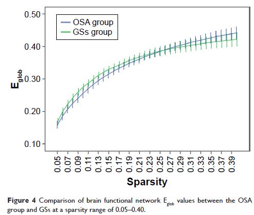
σ. The OSA group’s brain network showed significantly decreased Eglob (P <0.05) over the sparsity range
of 0.09–0.15, but significantly increased Eloc over the sparsity range of 0.23–0.40. In OSA patients, γ was
significantly negatively correlated with apnea–hypopnea index (AHI; r =−0.326, P =0.015) and
Epworth Sleepiness Scale (ESS; r =−0.274, P =0.043), λ was
significantly positively correlated with AHI (r =0.373, P =0.005) and ESS (r =0.269, P =0.047), and σ was
significantly negatively correlated with AHI (r =−0.363, P =0.007) and ESS (r =−0.295, P =0.029).
Conclusion: Our results suggest that the high degree of local integration and
integrity of the brain connections in OSA patients may be disrupted. The
topological alterations of small-world properties may be the mechanism of
cognitive impairment in OSA patients. In addition, σ, γ, and λ could be used as
a quantitative physiological index for auxiliary clinical diagnoses.
Keywords: obstructive sleep apnea, cognitive impairment, small-world, functional
MRI, topological properties