115195
论文已发表
注册即可获取德孚的最新动态
IF 收录期刊
- 3.6 Breast Cancer (Dove Med Press)
- 4.3 Clin Epidemiol
- 2.6 Cancer Manag Res
- 3.2 Infect Drug Resist
- 4.1 Clin Interv Aging
- 6.1 Drug Des Dev Ther
- 4.1 Int J Chronic Obstr
- 8.7 Int J Nanomed
- 2.5 Int J Women's Health
- 3.2 Neuropsych Dis Treat
- 2.4 OncoTargets Ther
- 2.6 Patient Prefer Adher
- 2.6 Ther Clin Risk Manag
- 3.1 J Pain Res
- 3.5 Diabet Metab Synd Ob
- 4.5 Psychol Res Behav Ma
- 3.4 Nat Sci Sleep
- 2.4 Pharmgenomics Pers Med
- 2.6 Risk Manag Healthc Policy
- 4.6 J Inflamm Res
- 2.3 Int J Gen Med
- 3.9 J Hepatocell Carcinoma
- 3.3 J Asthma Allergy
- 2.5 Clin Cosmet Investig Dermatol
- 3.0 J Multidiscip Healthc

成人广泛性焦虑症患者局部脑活动的自发改变
Authors Xia L, Li S, Wang T, Guo Y, Meng L, Feng Y, Cui Y, Wang F, Ma J, Jiang G
Received 3 February 2017
Accepted for publication 20 June 2017
Published 20 July 2017 Volume 2017:13 Pages 1957—1965
DOI https://doi.org/10.2147/NDT.S133853
Checked for plagiarism Yes
Review by Single-blind
Peer reviewers approved by Prof. Dr. Roumen Kirov
Peer reviewer comments 3
Editor who approved publication: Professor Wai Kwong Tang
Objective: We aimed
to examine how spontaneous brain activity might be related to the
pathophysiology of generalized anxiety disorder (GAD).
Patients and
methods: Using resting-state functional
MRI, we examined spontaneous regional brain activity in 31 GAD patients (mean
age, 36.87±9.16 years) and 36 healthy control participants (mean age,
39.53±8.83 years) matched for age, education, and sex from December 2014 to
October 2015. We performed a two-sample t -test on the
voxel-based analysis of the regional homogeneity (ReHo) maps. We used Pearson
correlation analysis to compare scores from the Hamilton Anxiety Rating Scale,
Hamilton Depression Rating Scale, State–Trait Anxiety Scale-Trait Scale, and
mean ReHo values.
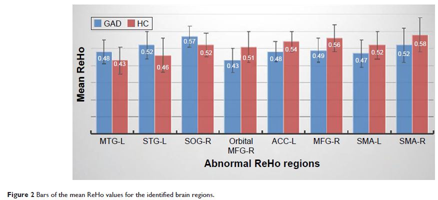
Results: We found abnormal spontaneous activity in multiple regions of brain in
GAD patients, especially in the sensorimotor cortex and emotional regions. GAD
patients showed decreased ReHo values in the right orbital middle frontal
gyrus, left anterior cingulate cortex, right middle frontal gyrus, and bilateral
supplementary motor areas, with increased ReHo values in the left middle
temporal gyrus, left superior temporal gyrus, and right superior occipital
gyrus. The ReHo value of the left middle temporal gyrus correlated positively
with the Hamilton Anxiety Rating Scale scores.
Conclusion: These results suggest that altered local synchronization of
spontaneous brain activity may be related to the pathophysiology of GAD.
Keywords: generalized anxiety disorder, functional magnetic resonance
imaging, resting state, regional homogeneity