115195
论文已发表
注册即可获取德孚的最新动态
IF 收录期刊
- 3.6 Breast Cancer (Dove Med Press)
- 4.3 Clin Epidemiol
- 2.6 Cancer Manag Res
- 3.2 Infect Drug Resist
- 4.1 Clin Interv Aging
- 6.1 Drug Des Dev Ther
- 4.1 Int J Chronic Obstr
- 8.7 Int J Nanomed
- 2.5 Int J Women's Health
- 3.2 Neuropsych Dis Treat
- 2.4 OncoTargets Ther
- 2.6 Patient Prefer Adher
- 2.6 Ther Clin Risk Manag
- 3.1 J Pain Res
- 3.5 Diabet Metab Synd Ob
- 4.5 Psychol Res Behav Ma
- 3.4 Nat Sci Sleep
- 2.4 Pharmgenomics Pers Med
- 2.6 Risk Manag Healthc Policy
- 4.6 J Inflamm Res
- 2.3 Int J Gen Med
- 3.9 J Hepatocell Carcinoma
- 3.3 J Asthma Allergy
- 2.5 Clin Cosmet Investig Dermatol
- 3.0 J Multidiscip Healthc

MRI assessment of whole-brain structural changes in aging
Authors Guo H, Siu W, D’Arcy RCN, Black SE, Grajauskas LA, Singh S, Zhang Y, Rockwood K, Song X
Received 12 April 2017
Accepted for publication 27 June 2017
Published 9 August 2017 Volume 2017:12 Pages 1251—1270
DOI https://doi.org/10.2147/CIA.S139515
Checked for plagiarism Yes
Review by Single-blind
Peer reviewers approved by Dr Amy Norman
Peer reviewer comments 3
Editor who approved publication: Dr Richard Walker
Purpose: One of the central features of brain aging is the accumulation of
multiple age-related structural changes, which occur heterogeneously in
individuals and can have immediate or potential clinical consequences. Each of
these deficits can coexist and interact, producing both independent and
additive impacts on brain health. Many of the changes can be visualized using
MRI. To collectively assess whole-brain structural changes, the MRI-based Brain
Atrophy and Lesion Index (BALI) has been developed. In this study, we validate
this whole-brain health assessment approach using several clinical MRI
examinations.
Materials and methods: Data came from three independent studies: the
Alzheimer’s Disease Neuroimaging Initiative Phase II (n=950; women =47.9%; age
=72.7±7.4 years); the National Alzheimer’s Coordinating Center (n=722; women
=55.1%; age =72.7±9.9 years); and the Tianjin Medical University General
Hospital Research database on older adults (n=170; women =60.0%; age =62.9±9.3
years). The 3.0-Tesla MRI scans were evaluated using the BALI rating scheme on
the basis of T1-weighted (T1WI), T2-weighted (T2WI), T2-weighted
fluid-attenuated inversion recovery (T2-FLAIR), and T2*-weighted
gradient-recalled echo (T2*GRE) images.
Results: Atrophy and lesion changes were commonly seen in
each MRI test. The BALI scores based on different sequences were highly
correlated (Spearman r 2>0.69; P <0.00001). They
were associated with age (r 2>0.29; P <0.00001) and
differed by cognitive status (χ 2>26.48, P <0.00001).
T2-FLAIR revealed a greater level of periventricular (χ 2=29.09) and deep
white matter (χ 2=26.65, P <0.001) lesions than others,
but missed revealing certain dilated perivascular spaces that were seen in T2WI
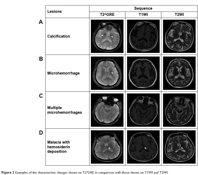
(P <0.001). Microhemorrhages
occurred in 15.3% of the sample examined and were detected using only T2*GRE.
Conclusion: The T1WI- and T2WI-based BALI evaluations
consistently identified the burden of aging and dementia-related decline of
structural brain health. Inclusion of additional MRI tests increased lesion
differentiation. Further research is to integrate MRI tests for a clinical tool
to aid the diagnosis and intervention of brain aging.
Keywords: aging, brain
atrophy and lesion index (BALI), brain health, MRI pulse sequences, structural
brain changes